Visualizing plant structure
Backlit pressed specimen
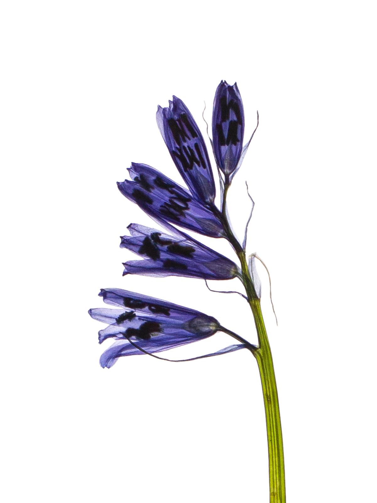
Botanical back-illumination is an imaging technique in which the light source is positioned behind a pressed plant specimen and directed toward the camera or scanner, realizable using a negative scanner, light table, or dedicated light box. Unlike conventional front-lighting, which records light reflected from the surface, back-illumination captures light transmitted through the tissue, absorbed variably by structural density and composition—denser cell walls and pigments blocking more light, while thinner veins and membranes transmit freely. The resulting images emphasize internal organization over surface appearance, with brightness and color variations revealing vascular systems and growth patterns clearly.
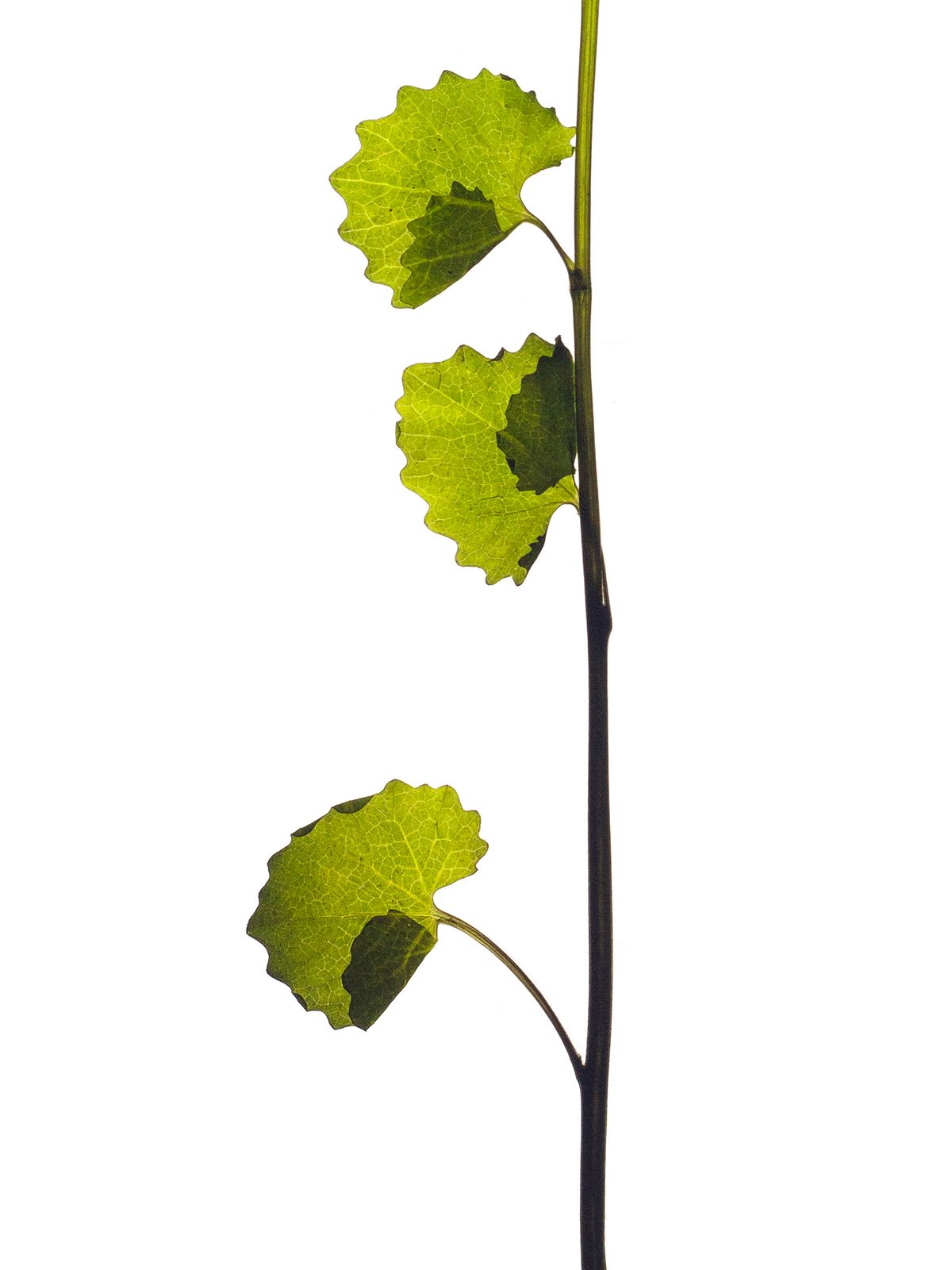
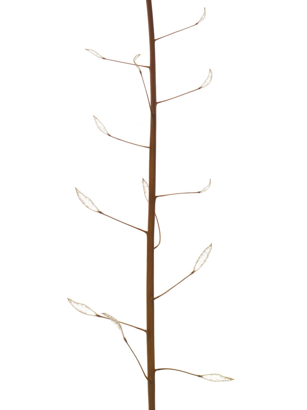
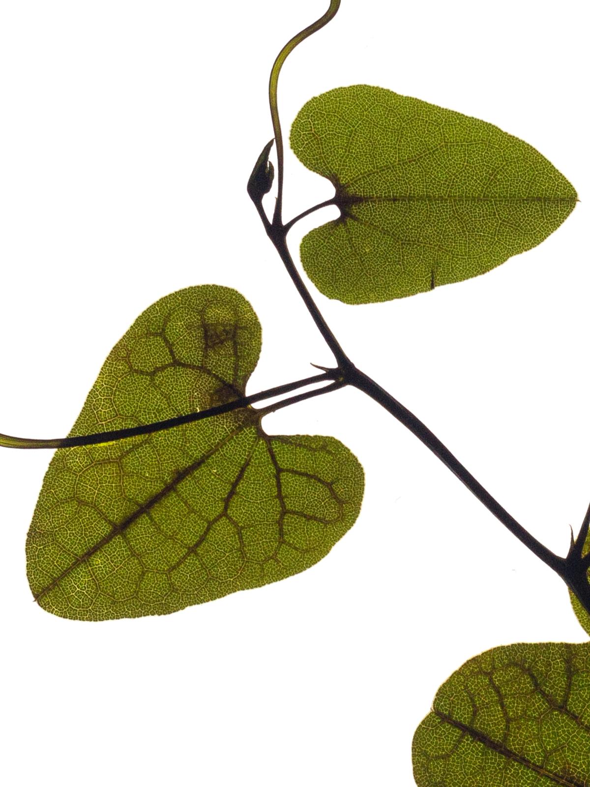
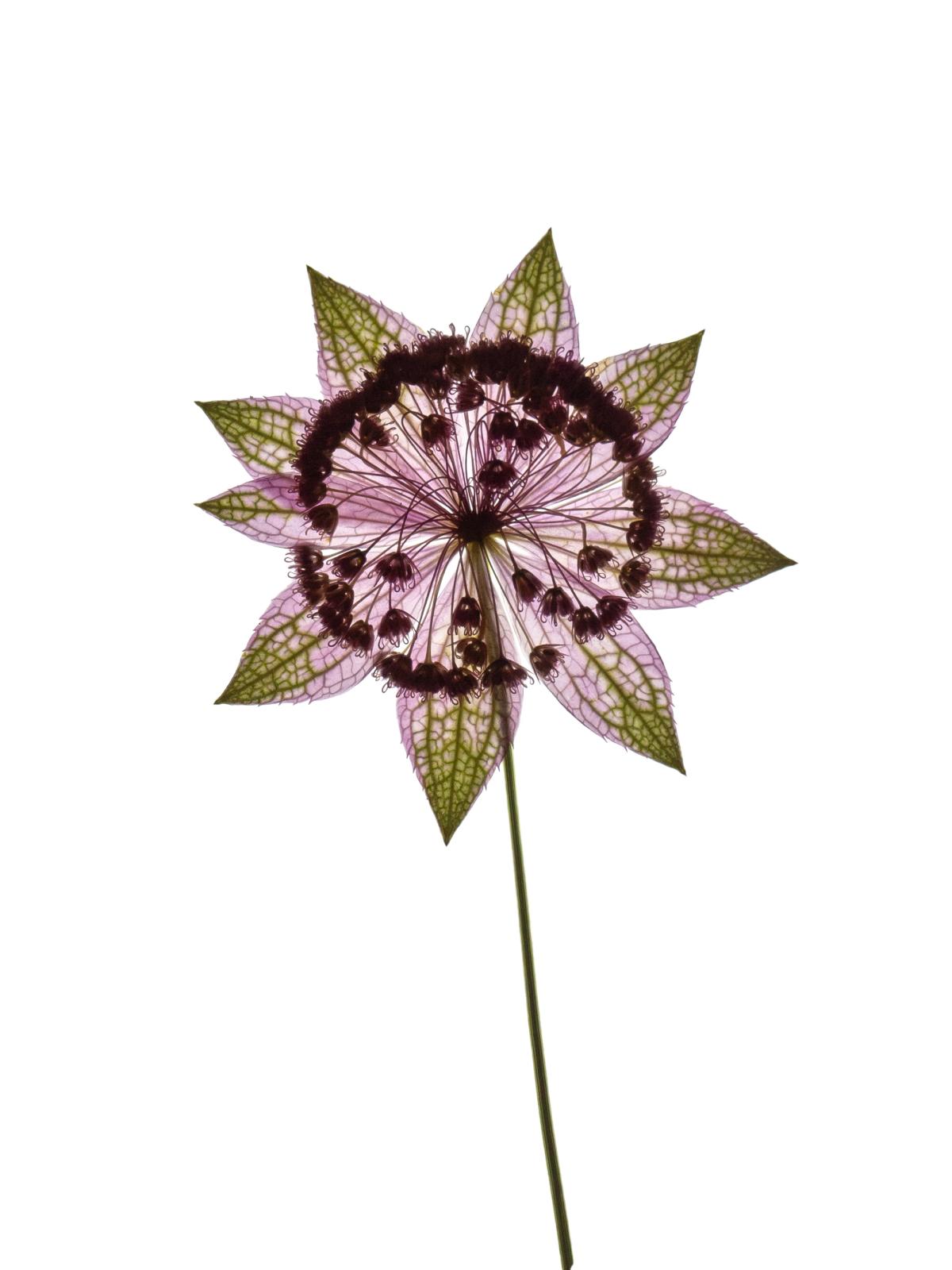
This technique excels in pressed specimens by highlighting translucence and layered details: in Alliaria petiolata, overlapping leaves glow with distinct depth, separating translucent lamina from shadowed edges; in Capsella bursa-pastoris, seed capsules become sharply visible. Similarly, Hyacinthoides non-scripta exposes delicate pistils and floral structures as fine, transmitted silhouettes, while Aristolochia baetica unveils the complex basal lobes and reticulate venation appear as luminous networks. In the backlit blossom of Astrantia maxima, the backview of florets becomes fully visible through the translucent petals and bracts.
By eliminating surface shadows and reflections, back-illumination renders repetition, branching, and symmetry—core principles of botanical growth—as precise forms, bridging scientific documentation and visual design in these examples.